
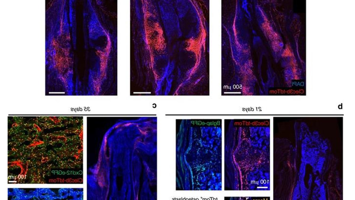
A study in Bone Research has overturned the prevailing belief that bone repair relies almost exclusively on cells already present in bone. Instead, the research identifies skeletal muscle as a hidden reservoir of progenitor cells that migrate to fracture sites and drive new bone formation. These cells, called fibroadipogenic progenitors (FAPs), convert into osteoblasts—the primary builders of healed bone—after injury, the team found.
Dr. Ugur M. Ayturk and colleagues at the Hospital for Special Surgery and Weill Cornell Medicine used genetic tracing in mice to track these cells. Under normal conditions, FAPs remain dormant in muscle, but following a fracture, they become a key contributor to healing. By day 21, nearly 28% of osteoblasts in the healing callus originated from muscle-derived cells, a proportion that may be even higher given the study’s tracking limitations.
This challenges decades of research that centered on periosteal cells and marrow stromal cells as the main drivers of bone repair. The new work shows muscle-resident FAPs play a far larger role. This demonstrated muscle, not the periosteum, supplies the majority of regenerative cells.
Muscle Cells Also Drive Unwanted Bone Growth
The findings also explain how these cells contribute to heterotopic ossification, the abnormal bone growth seen after severe trauma. Depleting these cells before BMP2 injection reduced ectopic bone volume significantly, but the effect reversed if depletion occurred after mineralization had already started, indicating other cell types could compensate.
The study reveals these cells have a dual role: aiding fracture healing while potentially driving unwanted bone growth in soft tissue. This duality creates a therapeutic opportunity, drugs could activate FAPs to speed healing or inhibit them to prevent heterotopic ossification, depending on timing and precision.
Previous research struggled to distinguish them due to overlapping genetic profiles. The cells remained dormant in healthy muscle but became active and bone-forming after injury.
The research also shows these cells behave differently depending on context. This suggests the molecular environment determines their fate, a detail critical for future treatments.
Read Also: UAE doctors urged to speed up breast cancer referrals
WNT Signaling and Bone Healing Targets Identified
Targeting these cells could address delayed unions, non-unions, and post-traumatic bone growth, offering a precise cellular target previously unknown.
Single-cell RNA sequencing revealed how these muscle-derived cells integrate into healing at a molecular level. Initially, they downregulated muscle-specific genes while upregulating mesenchymal progenitor markers like Postn and Col1a1. By day 14 post-fracture, a subset began expressing Spp1 (osteopontin) and Ibsp (bone sialoprotein), confirming their commitment to osteoblast lineage. The data also showed these cells contribute to marrow cavity reconstruction, adopting stromal identities such as Prx1 and Nes, indicating a broader role in restoring bone structure.
Mechanical cues further influenced their behavior. However, the study did not determine whether FAP activation alone could replicate these effects without mechanical stimulation.
Bone graft experiments reinforced muscle’s dominance in supplying regenerative cells. Even when the periosteum remained intact, most osteoblasts still traced back to muscle-resident FAPs. The persistence of labeled cells in both osteoblast and stromal populations, even in muscle-free grafts, suggests these progenitors may circulate systemically under certain conditions, though this was not explored further.
Timing Critical for Blocking Pathological Bone Growth
The study’s depletion experiments highlighted critical intervention windows in pathological bone growth. However, if depletion occurred after mineralization, typically by day 7, bone mass not only failed to decrease but increased, likely due to compensatory responses from other progenitor populations. This timing dependency may explain why some clinical trials targeting bone growth factors have produced inconsistent results.
After Achilles tendon severance, labeled cells appeared in mineralized deposits around the calcaneus and ankle joint, often via cartilage formation. This mirrors human cases where heterotopic ossification follows trauma, such as blast injuries or spinal cord injury, where abnormal bone forms in muscle rather than at fracture sites. The study did not determine whether these cells originate locally or migrate from distant sites, but their consistent presence in both fracture and ectopic models suggests a shared mechanism.


Leave a Reply